GSB-L868
$0.00 – $1,800.00Price range: $0.00 through $1,800.00
Our Cell Products
We offer a portfolio of high-quality, human iPSC-derived cell types. Each is validated for identity, functionality, and consistency—ideal for disease modeling, drug screening, electrophysiology, immunology, and tissue engineering.
Ventricular Cardiomyocytes


Human iPSC-derived Ventricular Cardiomyocytes
These cells are produced via standardized processes to possess a robust, ventricular-like phenotype. Their moderate spontaneous beat rate and well-defined action potentials make them particularly suitable for electrophysiological assays and compound profiling.
Benefits
- High purity ventricular phenotype
- Strong functional resemblance to human heart ventricular cardiomyocytes
- Reliable responses in electrophysiology, safety, and pharmacology assays
Product Specifications
|
Feature |
Detail |
|---|---|
|
Identity Markers |
≥ 85% cardiac Troponin T (cTnT+) by flow cytometry at Day 3; expression of ventricular marker MLC2v; sarcomeric markers (e.g. TNNT2), downregulation of progenitor/undifferentiated iPSC genes. |
|
Quantities |
≥ 1 × 10⁶ and ≥ 4 × 10⁶ viable cells |
|
Quality Control |
Cell count, viability, identity (FACS), functionality (e.g. Voltage Sensitive Dyes/Calcium Sensitive Probe: response to isoproterenol, dofetilide, nifedipine), mycoplasma testing |
|
Format |
Cryopreserved |
|
Donor & Reprogramming |
Specified donor (female/male; healthy/diseased as applicable); Sendai Virus reprogramming |
|
Shipping / Storage |
Dry shipper at -180°C to -135°C; store in vapor phase of liquid nitrogen |
|
Supplied with |
Cardiomyocyte culture medium; user-guide/instructions for thawing / seeding / maintenance / maturation |
Figure 1. Electrophysiological validation. Greenstone Biosciences ventricular cardiomyocytes baseline action potentials and response to hERG Channel blockade ( red symbols, 30nM, E4031).
Technical Data
- Clear structural sarcomere alignment, α-actinin / myosin heavy chain staining
- Action-potential recordings show fast upstroke, distinct plateau phase typical of human ventricular myocytes
- Ventricular CM APD>>Atrial CM APD
- Response to adrenergic stimulation (e.g. isoproterenol) as well as to ion channel blockers (e.g. L-type Ca²⁺ channel blockers, hERG blockers, Na channel blockers)
Atrial Cardiomyocytes
Human iPSC-derived Atrial Cardiomyocytes
Engineered to express atrial-specific electrophysiological and molecular signatures. They beat at spontaneous rates and have action potential morphologies characteristic of atrial cardiomyocytes—useful for atrial arrhythmia modeling and for testing drugs targeting atrial-selective pathways.
Benefits
- Atrial gene marker expression (e.g. KCNA5, NPPA, GIRK (IKAch))
- Electrophysiological profiles suited to atrial physiology studies
- Strong for comparative studies vs ventricular types; arrhythmia / AFib models
Product Specifications
Figure 2. Atrial hiPSC-CM electrophysiological validation. Atrial hiPSC-CM APD90 << Ventricular hiPSC-CM APD90. Greenstone atrial hiPSC-CMs respond to Dofetilide (100nM) as expected with significant APD prolongation.
- Identity: high purity atrial marker expression; minimal ventricular contamination
- Sizes, QC, format, shipping as per ventricular line
- Functionality: response to atrial-relevant ion channel modulators; beat rate and action potentials matching atrial reference data
- Response to adrenergic stimulation (e.g. isoproterenol) as well as to ion channel blockers (e.g. L-type Ca²⁺ channel blockers, hERG blockers, Na channel blockers)
- Atrial CM APD<<Ventricular CM APD
Fibroblasts
Human iPSC-derived Fibroblasts
Widely used to study ECM remodeling, fibrosis, cell signaling, and wound repair. These fibroblasts show robust secretion of matrix proteins and support for co-culture and 3D tissue systems.
Benefits
- High expression of fibroblast markers (e.g. Vimentin, FSP1, DDR2, PDGFR-α)
- Strong ECM production, contractility in gel contraction assays
- Compatible with co-cultures, organoids, 3D matrices
Product Specifications
Figure 3. Representative immunofluorescence images showing stage-specific cells express pluripotency genes (iPSC; NANOG and SSEA4), pass through epicardial cell lineage (EPC; ZO1 and WT1) and cardiac fibroblast (CF) genes (GATA4 and DDR2) during the differentiation process. DAPI=nucleus stain. Scale bars, 100µm. From Zhang et al, Methods Mol Biol. 2022;2454:109–115. doi: 10.1007/7651_2020_300
- Identity via marker panels; QC includes viability, mycoplasma, functional assays (e.g. ECM deposition)
- Supplied cryopreserved; sizes scaled to research or higher throughput needs
- Proper donor background info available
Smooth Muscle Cells
Human iPSC-derived Vascular Smooth Muscle Cells
Designed for vascular biology, these smooth muscle cells (SMCs) respond to vasoactive agents and display marker expression and phenotype switching characteristic of vascular SMCs.
Benefits
- Expression of key SMC markers (ACTA2, MYH11, CNN1, TAGLN)
- Functional contractile behavior; responsiveness to vasoconstrictors / dilators
- Useful for disease modeling (hypertension, vascular remodeling), tissue engineering, screening
Product Specifications
- Identity & purity assessed; QC includes contractility assays, viability, donor background
- Cryopreserved, multiple scale sizes; shipping & storage under liquid nitrogen vapor phase
Macrophages
Human iPSC-derived Macrophages
Tailored for immunology, inflammation, infectious disease, and cell-cell interaction studies. These macrophages can be polarized (M1 / M2), secrete cytokines, and perform phagocytosis reliably.
Benefits
- High expression of macrophage markers (CD14, CD68, CD11b)
- Functional assays: phagocytosis, cytokine profile upon stimulation (e.g. LPS, IFN‐γ)
- Good for modeling immune response, inflammatory disease, host-pathogen interaction
Product Specifications
- Identity/purity QC, functional validation (phagocytosis, polarization, cytokine secretion)
- Cryopreserved format; supply with macrophage media; standard donor info; mycoplasma free
Endothelial Cells
Human iPSC-derived Endothelial Cells
Human iPSC-derived endothelial cells provide a reliable and renewable source for vascular biology research. These cells exhibit robust endothelial identity, form functional vascular networks, and respond to angiogenic and inflammatory signals, making them a powerful platform for disease modeling, drug discovery, and regenerative medicine.
Benefits
- Expression of key endothelial markers (CD31/PECAM1, VE-Cadherin, vWF, eNOS)
- Responsive to shear stress, cytokines, and pro-angiogenic/anti-angiogenic factors
- Suitable for vascular disease modeling (atherosclerosis, angiogenesis, thrombosis), tissue engineering, and compound screening
Product Specifications
|
Feature |
Detail |
|---|---|
|
Identity Markers |
Sucrase isomaltase (enterocytes), (MUC2 (goblet cells), LGR5 (stem cells), Lyz (Paneth cells) |
|
Quantities |
≥ 500 organoids/ crypt like structures per vial |
|
Quality Control |
Markers characterization , mycoplasma testing |
|
Format |
Cryopreserved |
|
Donor & Reprogramming |
Healthy/ diseased donor-derived iPSCs; Sendai virus reprogramming |
|
Supplied with |
Handling guide and instructions for differentiation and culture |
- Identity & purity assessed; QC includes tube formation, nitric oxide production, and viability assays
- Cryopreserved, multiple scale sizes available; shipping & storage under liquid nitrogen vapor phase
Epithelial Cells
Human iPSC-derived Intestinal Epithelial Cells
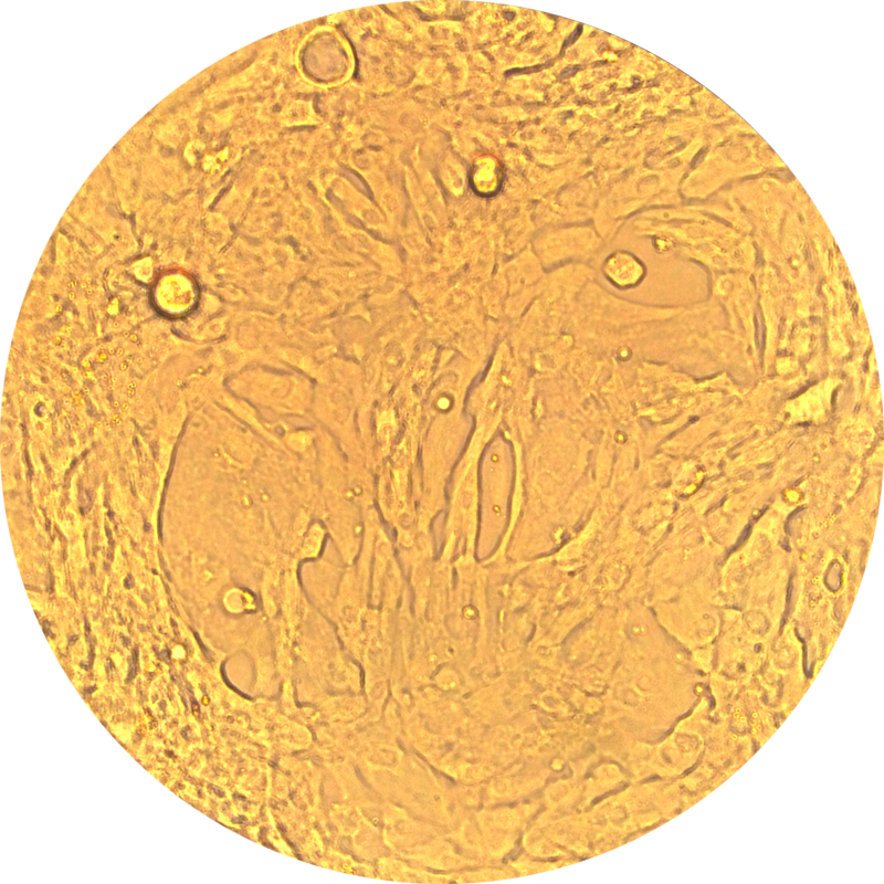
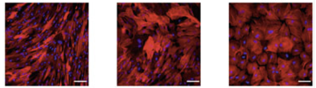
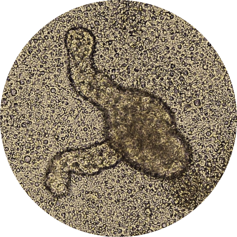
These organoids mimic the structure and cellular diversity of human intestinal epithelium.IPSC-derived intestinal products show a crypt-like structure, which is the common feature of intestinal epithelium. They contain enterocytes, goblet cells, Paneth cells, and enteroendocrine cells. They are valuable tools for studying gut barrier function, host-microbiome interactions, screening drugs, assessing pharmacokinetic profiles of potential medications, and testing the toxicology of possible interventions.
Benefits
- Physiological 3D intestinal architecture
- Contains absorptive and secretory cells
- Suitable New Approach Method (NAM) for gut disease modeling
- Develops a high-throughput platform for drug screening and assessing the toxicology of potential drugs.
Product Specifications
|
Feature |
Detail |
|---|---|
|
Identity Markers |
Sucrase isomaltase (enterocytes), (MUC2 (goblet cells), LGR5 (stem cells), Lyz (Paneth cells) |
|
Quantities |
≥ 500 organoids/ crypt like structures per vial |
|
Quality Control |
Markers characterization , mycoplasma testing |
|
Format |
Cryopreserved |
|
Donor & Reprogramming |
Healthy/ diseased donor-derived iPSCs; Sendai virus reprogramming |
|
Supplied with |
Handling guide and instructions for differentiation and culture |
